What is vitrectomy?

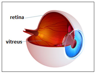
Vitrectomy; The thin nerve layer that surrounds the inner surface of the eye like a sheet and creates visual signals is called the retina. The gel substance that fills the inside of the eye is called the vitreous.
The vitreous is normally attached to the retina. In some diseases, the vitreous substance becomes cloudy and can impair vision. Sometimes the vitreous needs to be removed to interfere with the retina.
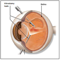
The surgery to remove and clean the vitreous is called vitrectomy. In this way, the following necessary interventions can be made:
- Clearing the hemorrhage
- Correction of scar tissue and wrinkle
- Repairing the macular hole
- Repairing the retinal detachment
- Removal of intraocular foreign body
- In which cases is vitrectomy surgery required?
Vitrectomy is generally performed in cases where the vitreous (gel substance) becomes cloudy and in some retinal diseases. These are diseases such as:
- retinal detachment
- macular hole
- diabetes-related hemorrhage and retinal diseases
- epiretinal membrane
- complications related to cataract surgery
- sampling for diagnostic purposes
- trauma and intraocular foreign body
- endophthalmitis (intraocular infection)
- vitreous haze due to uveitis
How is vitrectomy surgery performed?
Vitrectomy surgery is an outpatient procedure. So the patient does not need to be hospitalized. If there is no special case, the patient is discharged on the same day. In most patients, regional (local) anesthesia applied to the corner of the eye is sufficient.
General anesthesia can be applied in pediatric patients, panic attack patients, eye traumas or operations that are expected to take a very long time.

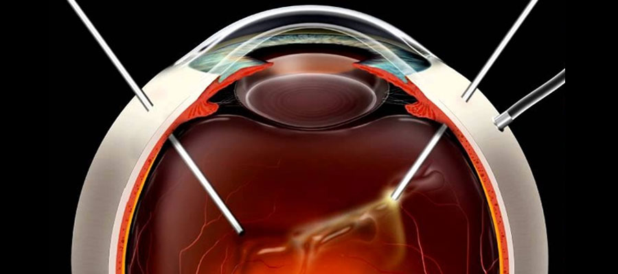
Before starting the operation, the eye is wiped and covered in a sterile manner. Then, with very thin instruments, 3 incisions of approximately 1 mm are made in the white part of the eye just behind the pupil and the operation is started.
At first, the vitreous gel is removed. Then, the treatment of diseased tissues such as detachments, holes, membranes in the retina is performed. If necessary, laser treatment is applied. At the end of the surgery, liquid, air, gas or silicone is left in the eye.
The fluid produced by the eye itself is replaced by the substance left in the eye over time. This is 1 week for air, 2-8 weeks for gas. Silicone, on the other hand, does not disappear in the eye and requires a second surgery to be removed.
What are the advantages and disadvantages of putting gas or silicone into the eye?
The gas put into the eye is resorbed over time. So it is not necessary to take it with a second surgery. But as long as there is gas in the eye, the vision will be blurry. After the gas disappears, the vision improves.
Gas is not preferred for patients who need early vision gain. Additionally, when gas is put in, it will be necessary to stand in a certain head position according to the location of the disease in the retina.
Gas is not preferred in patients with diseases such as spinal-cervical hernia and who cannot maintain the head position. Additionally, a person with gas in his eye cannot go to high altitude and cannot get on the plane until the gas disappears.
Silicone, on the other hand, does not impair vision because it is transparent, you can get on a plane, no head position is required, and it can be left in the eye for a long time. Silicone is preferred in some serious pathologies or people who cannot maintain the head position.
However, the silicone must be surgically removed after the healing process. This is an extra trouble. If it is left in the eye for a long time, its structure will deteriorate over time and it can cause eye pressure. It may damage the lens and cornea.
What are the risks of vitrectomy surgery?
Today, it is a surgery with a high success rate and few complications. However, it does involve certain risks. The most common complication is the development of cataract.
Additionally, complications such as bleeding, infection, increased intraocular pressure, recurrence of the disease, retinal detachment, and light damage to the retina are observed. But these are always rare problems.


