Floaters, Flashes of Light (Photopsia) and Posterior Vitreous Detachment
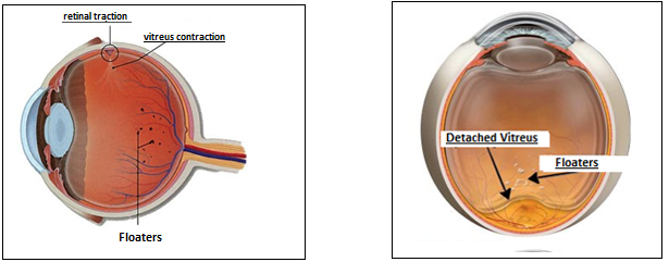
The gel substance that fills the inside of the eye is called the vitreous. The vitreous is normally attached to the retina. With increasing age, the gel substance liquefies and shrinks. When these changes reach a certain stage, the gel substance begins to separate from the retina. As the vitreous separates, it makes retraction on the retina from the places where it is tightly adhered.
With the effect of this traction, nerve cells are stimulated and flashes of light, which are more evident in the dim-lit environment, are seen. This detachment process is usually completed in a few months without any problems.
As a result of these changes, the gel substance concentrates in the eye and the fibers that make up the gel are clustered together. The clustered fibers float inside the eye and cast a shadow on the retina. They look like flying flies or small floating spots, especially in bright light or when looking at a white ground

Who is at risk?
Posterior vitreous detachment is a natural phenomenon and part of the aging process seen in 75% of people over the age of 65. It is rare under the age of 40. However, it can also be seen at an early age with some accompanying diseases. These diseases are as follows:
- High myopia
- After cataract surgery
- Eye traumas
- Uveitis (eye inflammation)
What are the symptoms of posterior vitreous detachment?
Small floaters happen to most people and are considered normal. Sudden increase in floaters is the first sign of posterior vitreous detachment. Floaters usually look like flies, spider webs, or tulle curtains. Sometimes these floaters may be accompanied by flashes of light.
Flashes of light are more noticeable in dim conditions and are in the outer parts of the visual field. It’s like a lightning flash or a fluorescent lamp flashing. It occurs due to the stimulation of nerve cells as the gel layer detaches from the retina. These events can last from a few weeks to several months.
When detaching the vitreous, it can sometimes rupture a small vein and cause intraocular bleeding. In this case, a more dense curtain-shaped shading occurs.
Does posterior vitreous detachment threaten vision?
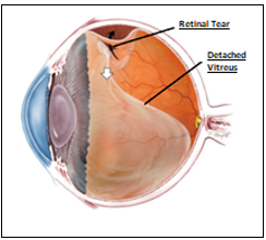
Posterior vitreous detachment is a harmless process in most people and is completed without affecting vision. Floaters also decrease over time. However, sometimes as the vitreous detaches, it can cause a tear in the retina and retinal veins.
Tears of retinal veins causes intraocular hemorrhage and may result in decreased vision. This type of bleeding usually clears up on its own within a few months.
Retinal tears is a condition that requires urgent intervention. If it is left untreated, it can develop into a more serious disease called retinal detachment.
How is posterior vitreous detachment diagnosed?
After the routine eye examination, the pupils are dilated with drops and the back of the eye is examined with special lenses. Posterior vitreous detachment is usually diagnosed with this examination.
Since the vitreous is a transparent layer, sometimes detachment may not be seen on examination. In this case, your doctor may request one of the eye ultrasound or eye tomography (optical coherence tomography) examinations.
How is posterior vitreous detachment treated?
If posterior vitreous detachment is not accompanied by an additional problem, no treatment is required. Floaters will automatically decrease. In some patients, these floaters do not disappear and if they are located too close to the visual field, they may cause discomfort to the patient.
In this case, the fibers causing the floaters can be cleaned with vitrectomy surgery. All risks should be explained to the patient before the decision of surgical operation.
If a retinal tear is detected during the examination in a patient with posterior vitreous detachment, laser treatment is immediately performed around the tear.
If there is no tear in the retina, the patient is informed and followed up. Until the vitreous detachment process is completed and the flashes of light are over, the patient is called for controls again for a few months and examined


