What Is Retinal Vein Occlusion?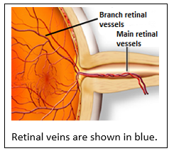
Retinal Vein Occlusion; The thin nerve layer that surrounds the inner surface of the eye like a sheet and creates visual signals is called the retina. Nutrition of the retina is provided by the retinal vascular system.
The veins that take blood from the retina and carry it back to the body’s vascular system are called retinal veins. The occlusions that occur in these veins are called retinal vein occlusion.
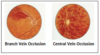
If the occlusion occurs in the main vein, it is called ‘central retinal vein occlusion’ and if it occurs in the vein branches, it is called ‘branch retinal vein occlusion’.
What are the causes of retinal vein occlusions?
The slowing of blood flow in retinal veins can cause thrombus formation and vein occlusion. Thrombus formation occurs due to reasons such as impaired blood flow, excessive blood coagulation, and damage to the inner surface of the blood vessel.
Some underlying diseases may be risk factors for these events. These are hypertension, diabetes, blood coagulation disorders, glaucoma (eye pressure) and cardiovascular diseases.
Your ophthalmologist may request an evaluation from the internal medicine or cardiology department in terms of coagulation disorders and cardiovascular diseases. If an underlying disease is detected, treatment must be provided.
What are the symptoms of retinal vein occlusions?
The most common symptom of retinal vein occlusion is painless loss of vision in one eye. While vision loss is almost always present in central retinal vein occlusions, there may be dark vision only in a certain area in branch occlusion.
Additionally, if the occluded branch is far from the visual center, it may not give any symptoms. In such unrecognized and untreated cases, there may be floaters or sudden vision loss due to intraocular bleeding after months.
Do retinal vein occlusions give any symptoms beforehand?
Central retinal vein occlusions can sometimes be preceded by blackouts that come and go. These are called temporary blackouts. Subsequently, vein occlusion may develop.
How is retinal vein occlusion diagnosed?
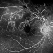
Angiographic appearance in branch occlusion
After the routine eye examination, the pupils are dilated with drops and the back of the eye is examined with special lenses.
In the examination, the condition of the retinal veins, the presence of hemorrhages, and the presence of fluid accumulation (edema) in the visual center are evaluated. According to the examination findings, your doctor may request eye angiography and/or eye tomography (OCT-optical coherence tomography) examinations.
Eye angiography helps to identify obstructed and damaged veins, undernourished retinal areas and abnormal new veins, if any. Eye tomography is used to determine the amount of fluid accumulation in the visual center (macula). This measurement is also followed for the response to treatment.
What is the treatment for retinal vein occlusions?
Treatment of retinal vein occlusions begins with the identification and treatment of the underlying risk factors. Control of blood pressure, lowering of cholesterol levels and treatment of coagulation disorder, if present, should be done. Otherwise, re-occlusion or occlusion in the other eye may occur.
Treatment of retinal vein occlusions is not to open the vein occlusion, but to correct the side effects caused by the occlusion. The most common cause of vision loss in this disease is fluid accumulation in the macula, which is the visual center of the retina, namely macular edema.
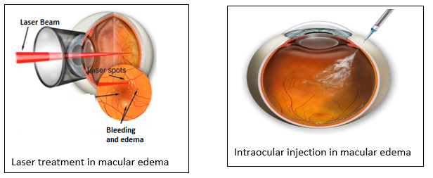
Intraocular injections are the most common treatment for macular edema. The drugs given intraocularly prevent the vessels from leaking and the edema heals. Usually, more than one injection is required at regular intervals.
There are several different types of drugs available today. The drug to be applied is determined by the condition of the eye and the preference of the physician. In branch vein occlusions, unlike main vein occlusions, edema may be small and localized. In this case, laser treatment can be applied to the edematous area.
The increase in vision with laser is slow and can take months. The disadvantage of intraocular injections is that the effect is temporary and multiple applications are required. The fact of how many and how often injections will be required becomes only evident during follow-up and varies from patient to patient.
Another undesirable side effect of retinal vein occlusion is abnormal new vein formation in the retina. These veins are much more fragile than normal veins and may cause vision loss by bleeding into the eye.
In addition to edema, laser is also used in the treatment of new vein formations. Laser burns are applied to retinal areas with impaired blood supply in a few sessions. Thus, new vessel formations are destroyed. Intraocular bleeding, that is, the risk of surgery and blindness, is prevented.
Despite all the treatments, sometimes the patient may not realize the situation and the patient may come with intraocular bleeding. In this case, the only treatment option is vitrectomy surgery. In vitrectomy surgery, very small holes are made in the eye and the eye is entered. Intraocular bleeding and membranes on the retinal surface are cleaned. Laser is applied to retinal areas with impaired blood supply.
If these treatments are not applied in a timely manner, vein formations may envelop the entire eye, causing serious eye pressure that will lead to blindness and even removal of the eye.


